Part 1: decoding eagle rays in the lab
Like all work in the field of Molecular Ecology, our Save Our Seas Foundation funded eagle ray research comprises three broad components:
- Fieldwork,
- Labwork, and
- Bioinformatics (data analysis)
Our first blog gave some background about the field of Molecular Ecology and our second detailed the fieldwork our team has completed to collect tissue samples from eagle rays for kinship analysis and CKMR. This entry is Part I in a two-part blog describing the critical labwork that follows tissue collection and precedes data analysis and inference.
Molecular Ecology labwork is a funny thing. From one perspective it’s very simple: about 75% of our labwork entails simply moving (very small amounts of) liquid from one tube to another. Another 15% or so of the labwork involves shaking, spinning, and heating up/cooling down those tubes. Hardly the kind of tasks that require a PhD; a toddler could shake, spin, and heat up (and probably ingest) tubes of liquid just as easily. However, the biochemical processes that occur when liquids with DNA are combined, mixed, and heated/cooled are quite sensitive and fascinating.
Ultimately, the goal of our labwork is to turn tissue samples into data. For our eagle ray research, the first step in the lab is to extract DNA from the tissue samples that were collected in the field. Nearly every cell in a living organism contains DNA – a.k.a. “the code of life” – within a nucleus that resides at the cell’s core. Accessing the DNA requires us to first break down the barriers surrounding the individual cells (the cell membrane) and then break down the barrier surrounding the nucleus (the nuclear membrane) to expose the DNA contained therein. We do this using an intrepid enzyme called “proteinase K”. The primary function of Proteinase K (also known as PK) is to break down other proteins, such as those that constitute the cellular and nuclear membranes of a cell.
We mix each tissue sample with some PK, heat it up (to promote enzyme activity), and leave it overnight, which gives the PK time to do its work. The next day, we remove the homogenous mixture and extract the DNA from the mixture using microscopic magnetic beads that bind only to DNA. Everything else – all the residual proteins and cellular components – is washed away using various wash solutions so that only pure DNA suspended in water remains.
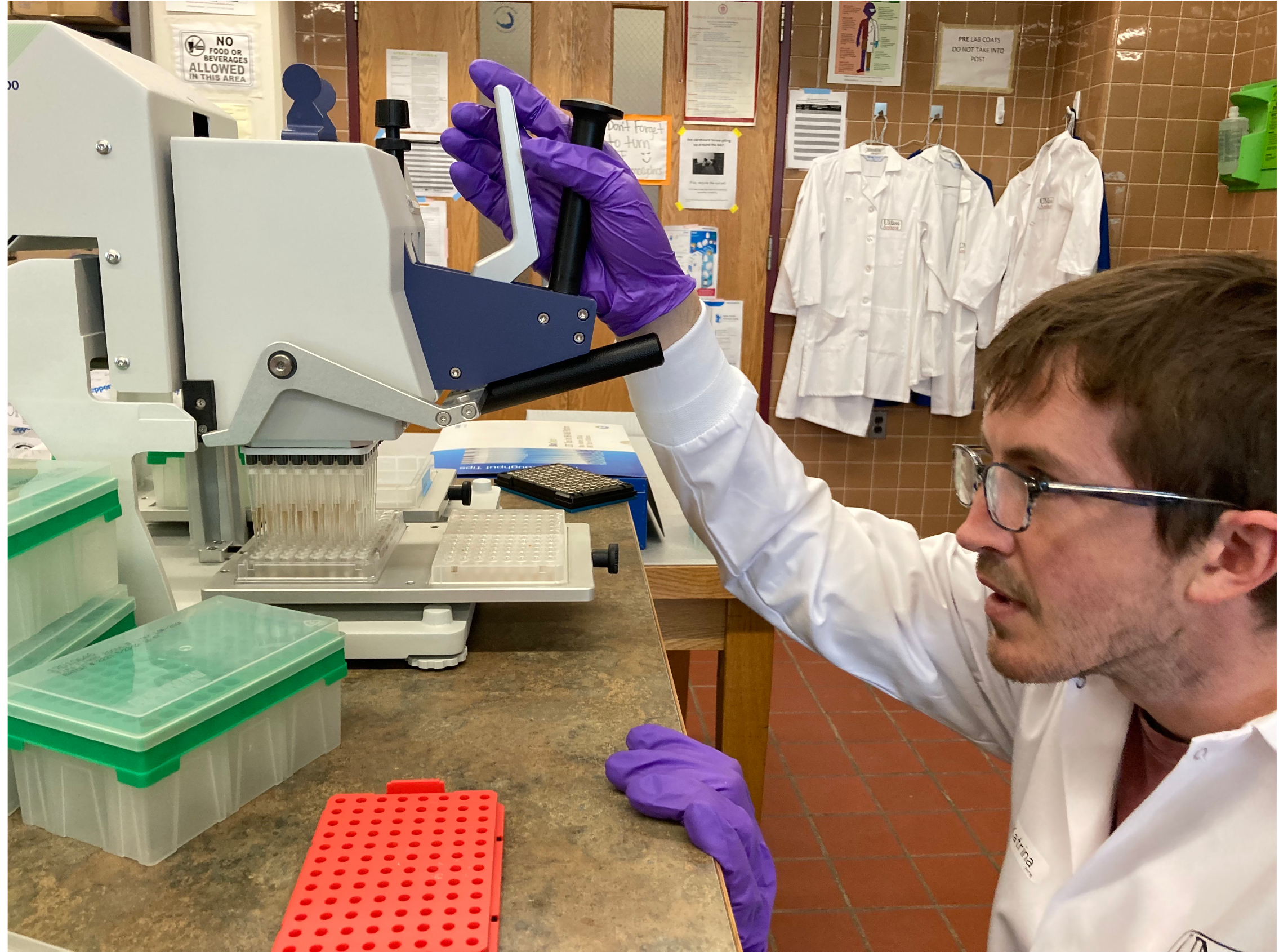
Extracting DNA from 96 tissue samples simultaneously using a high-throughput tool called a “Liquidator”. Photo © Katrina Phillips.
Once we’ve isolated the DNA, we check to ensure that it has not undergone significant damage or degradation; if it has, then the information contained in the DNA will likely be lost during subsequent processing steps. The process of checking DNA quality and quantity is quite fascinating! DNA is too small to be observed with the naked eye, so we cannot just look at our tube of DNA to check whether it’s intact; rather, we indirectly infer DNA integrity using methods that take advantage of its polarity (i.e., electromagnetic charge) and/or its absorbance at different wavelengths of light. For example, by adding DNA to a porous agarose gel and applying an electrical current, we can infer the size of DNA molecules based on how long it takes for the current to draw the DNA from one side of the gel to the other: a longer “migration time” means the DNA had a harder time maneuvering through the miniscule pores of the gel. This means that the DNA exists in long fragments that are less maneuverable, which in turn signifies that the DNA is intact. Meanwhile, the presence of contaminants within our DNA can be detected via light absorbance. Pure DNA absorbs light at specific wavelengths: if we expose the DNA to known wavelengths of light, we can quantify which wavelengths are absorbed by the DNA solution. If our DNA solution absorbs light at wavelengths that vary from our expectations, then it is likely that the DNA is contaminated with other substances (e.g., proteins, salts). In such cases, there are various steps that we can take to try to remove the contaminants and purify our DNA. Once we have high quality and pure DNA, then the truly bonkers labwork begins, which we will cover in Part II of this blog.