
Wrap your head around smalltooth sawfish neuroanatomy
The Yopak ZoMBiE Lab at the University of North Carolina Wilmington (UNCW) has been hard at work developing and optimising protocols using magnetic resonance imaging (MRI) to non-destructively characterise neuroanatomy of the smalltooth sawfish, Pristis pectinata (check out our previous blog post to learn more!).
Along with Dr. Kara Yopak, Dr. Emily Peele, and our talented team of collaborators, we’ve recruited PhD student Maria Pierce to take on this project. Maria has not only hit the ground running with starting a graduate program at UNCW, but also with getting trained on MRI methodology and neuroanatomy to begin initial data collection.
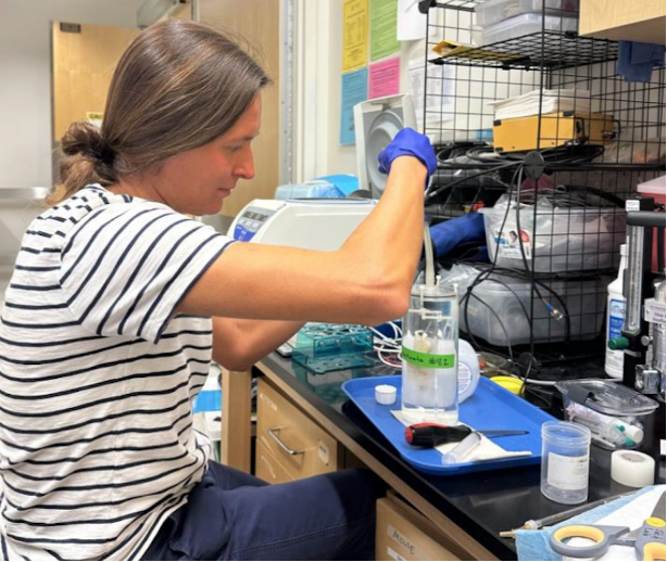
Maria Pierce preparing a brain sample for MRI. Photo © E. Peele
Lessons in neuroanatomy
As a new PhD student in the lab, the first step in building a brain atlas for P. pectinata is familiarising yourself with elasmobranch (shark, skates, and rays) neuroanatomy and sensory structures (such as the eyes, nose, ears). The brain plan of elasmobranchs, with distinct forebrain, midbrain, and hindbrain regions, has a “blueprint” that is shared across nearly all vertebrates. If you’ve never taken a neurobiology course before, the brain can be divided into six major brain regions (olfactory bulbs, telencephalon, mesencephalon, diencephalon, cerebellum, and medulla oblongata) that can all be identified whether you are a shark, lizard, or a primate. Yet, within this shared framework, elasmobranchs exhibit a lot of diversity in brain size and outlook – with expansion of brain regions that often correlates with sensory specialisations and behaviours (check out our first blog post to learn more!).
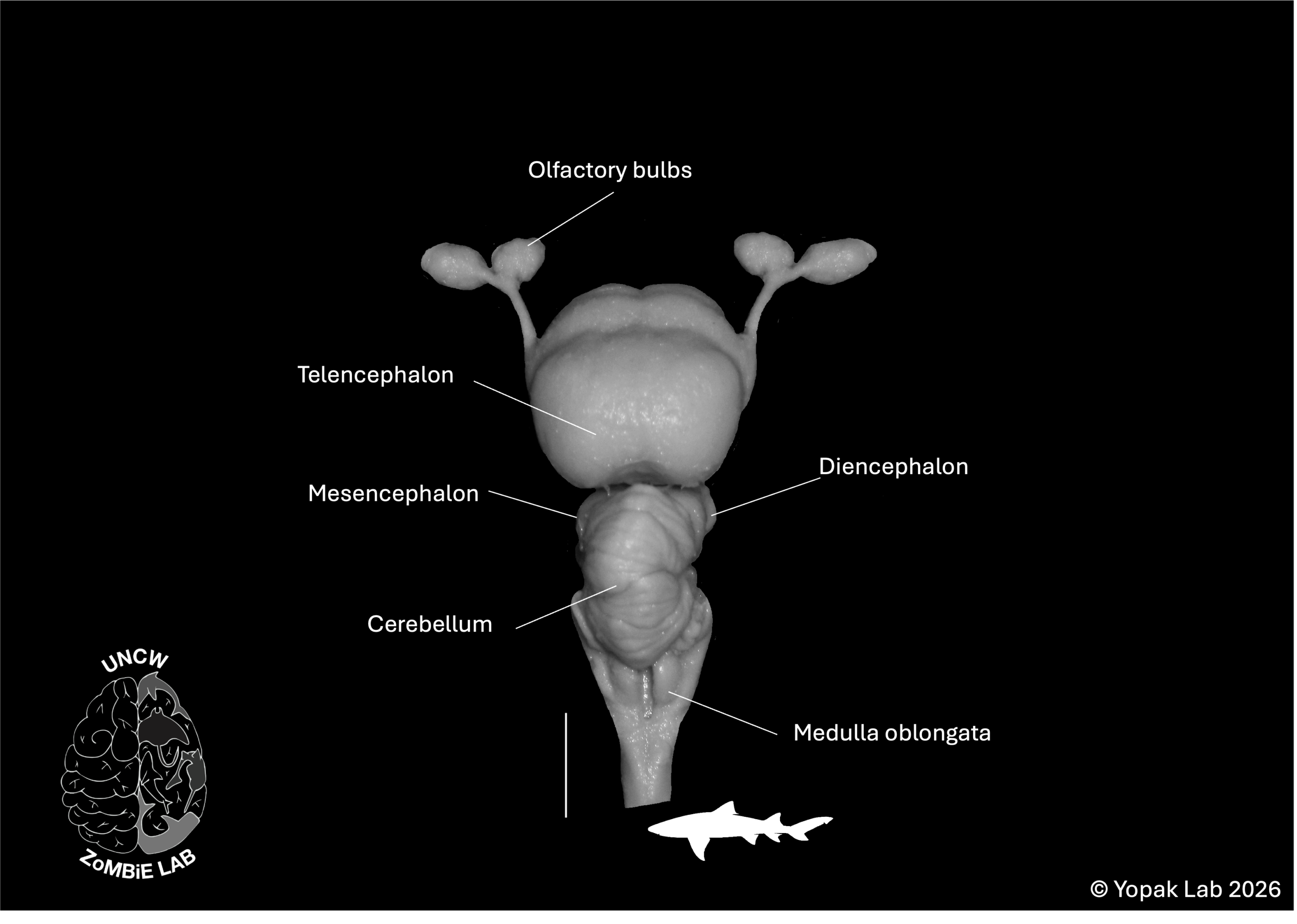
Want a quick neuroanatomy lesson? The figure above is showing the brain of the lemon shark (Negaprion brevirostris), pointing out the six major brain regions that can be identified in elasmobranchs (and nearly all vertebrates!) Scale bar = 1cm. Figure adapted from Yopak (2022)
The ability to examine the variability in the relative size of these major brain regions across species and throughout life within a single species allows us to explore the relationships between brain morphology, behaviour, and sensory ecology. Moreover, the opportunity to characterise the brain morphology of such a rare and Endangered species like P. pectinata could fill critical gaps in our understanding of vertebrate brain evolution and support more integrative assessments of ecological resilience and vulnerability.
Because P. pectinata are so rare, we are examining variation in the brain in this Critically Endangered species by acquiring MRI scans and characterising size and structure of those six brain regions – in other words, we are dissecting their brains without using a scalpel. We aim to look at if (and how) the brain changes throughout life in this species, as well as whether we can identify neural anomalies in specimens from recent mortality events in South Florida.
A project such as this requires collaboration across a massive team of amazing researchers. In addition to support from SOSF, this work is bringing together FWC, the Fish and Wildlife Foundation of Florida, Havenworth Coastal Conservation, and the Bonefish Tarpon Trust
Want to read more about some of our lab’s work discussed above? Check out our publications page!
**Reference:
Yopak KE. 2022. Advances in Chondrichthyan Neurobiology. In Biology of Sharks and Their Relatives, Edition 3. New York: CRC Press. pp 105-142 ISBN 9780367861179